
Surgery is all about precision. The more a surgeon knows before making the first incision, the better outcome a patient receives. This is where preoperative risk assessment comes in — it’s the process of evaluating potential complications before surgery even begins.
Importance of preoperative risk assessment
Every surgery carries risks. Blood loss, organ damage, unexpected anatomical variations — there’s a long list of potential complications. Preoperative risk assessment involves evaluating the patient’s overall health, reviewing medical history, and analyzing imaging data to understand the surgical site.
The goal? To identify these risks before surgery to improve patient safety and optimize outcomes. This is where 3D anatomy models play a crucial role. By providing highly detailed, patient-specific visualizations, they help surgeons detect potential complications early, refine their surgical approach, and enhance overall precision.
Limitations of traditional risk assessment methods
Traditional risk assessment methods have long been the foundation of surgical planning, but they come with significant limitations:
Flat 2D representations
Imaging techniques like X-rays, CT scans, and MRIs present a flat, two-dimensional view of complex anatomical structures. This makes it challenging to fully grasp the depth and spatial relationships between organs, blood vessels, and bones.
Interpretation challenges
Even the most experienced surgeons may struggle to mentally reconstruct a three-dimensional structure from multiple 2D image slices. This can lead to misinterpretations, which increases the risk of complications during surgery.
Lack of interactivity
Traditional imaging methods can offer the opportunity for direct manipulation or interaction. A surgeon can only view the scans from predetermined angles, limiting their ability to explore different perspectives and anticipate potential complications before surgery.
How 3D anatomy models enhance risk assessment
Custom 3D anatomy models revolutionize risk assessment by providing surgeons with highly detailed, patient-specific visualizations. Here’s how they enable better surgical planning:
Improved visualization and spatial understanding
One of the biggest advantages of 3D anatomy models is how they transform medical imaging into interactive, three-dimensional representations. Instead of trying to piece together a patient’s anatomy from multiple 2D slices, surgeons can examine a fully formed, life-like model. What are the outcomes?
- More accurate assessment: Seeing the anatomy in 3D helps surgeons better understand spatial relationships, for instance, the proximity of blood vessels to a tumor.
- Early identification of risks: Variations in anatomy that might be missed in 2D images become clear in 3D models.
- Virtual exploration: Surgeons can rotate and zoom in on digital models to study every detail before stepping into the operating room.
Personalized surgical planning
No patients are alike as our anatomy can vary significantly. A 3D anatomy model offers a customized roadmap for surgery, allowing for more precise planning.
- Tailored surgical approach: Knowing the exact structure of a patient’s anatomy helps surgeons choose the safest, most effective techniques.
- Reduced surprises: Understanding anatomical anomalies ahead of time means fewer unexpected complications during surgery.
- Minimized invasiveness: More precise planning can lead to less invasive procedures, reducing recovery times and improving patient outcomes.
Enhanced communication and collaboration
Surgery isn’t a solo act; it’s a team effort that involves surgeons, anesthesiologists, radiologists, and other specialists. 3D models improve communication among medical professionals and with patients alike.
- Better teamwork: Surgeons and specialists can collaborate more effectively when they’re looking at the same detailed, three-dimensional representation.
- Clearer patient discussions: Showing a patient their own 3D anatomy helps them understand their condition and the planned procedure, leading to more informed consent.
- More confidence for everyone: When medical teams and patients are on the same page, outcomes improve and anxiety decreases.
How surgeons can get custom 3D anatomy models 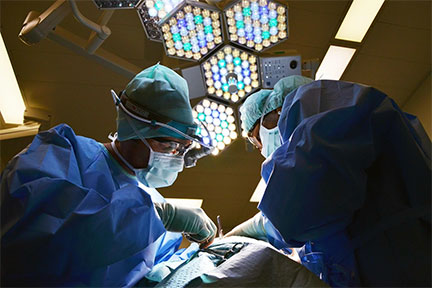
Surgeons can create patient-specific 3D anatomy models using specialized software that transforms standard medical images into interactive 3D visuals. For instance, advanced imaging software like OsiriX allows seamless import of CT scans and uses AI features to generate highly detailed models for surgical planning. Or they can use InVesalius, an open-source tool that not only reconstructs 3D models from CT or MRI scans but also allows exporting files compatible with 3D printing.
For highly complex cases or custom models requiring advanced precision, surgeons can also turn to specialized companies that offer professional 3D modeling services. Such companies have experienced 3D modelers and use cutting-edge software to produce scientifically accurate, patient-specific models tailored for surgical planning.
Final word
Custom 3D anatomy models are changing the game in preoperative risk assessment. By providing detailed, patient-specific representations of anatomy, they help surgeons visualize, plan, and execute procedures with greater accuracy and confidence.
As technology continues to advance, we can expect even more innovations in 3D modeling, including AI-enhanced models and virtual reality integration. For now, one thing is clear: the future of surgical planning is three-dimensional, and it’s making surgery safer, smarter, and more effective than ever.